Shoulder Joint Anatomy Diagram. Just remember the articulating surfaces. Moderate to serve pain along the outer edge of the clavicle, when raising arm over the head, and/or when reaching arm across the body. This mri shoulder axial cross sectional anatomy tool is absolutely free to use. Start studying shoulder joint anatomy.
In common usage, shoulder joint mostly refers to the glenohumeral joint, the major joint of the shoulder but can also include acromioclavicular joint. 8 name the arteries and the. Use the mouse scroll wheel to move the images up and down alternatively use the tiny arrows (>>) on both side of the image to move the images. This incongruent bony anatomy allows for the wide range of movement available at the shoulder joint but is also the reason for the lack of joint stability. Home > blog > anatomy > shoulder anatomy: The human shoulder is the most mobile joint in the body. We used a 3d volume. Simple easy notes for quick revision for exams. The glenohumearal joint has a greater range of motion than any other joint in the body. Learn vocabulary, terms and more with flashcards, games and other study tools.
The shoulder joint is vulnerable to dislocations from sudden jerks of the arm, especially in children before strong muscles have developed.
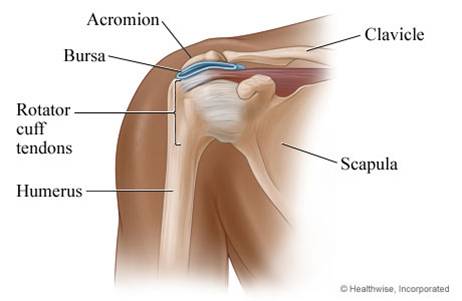
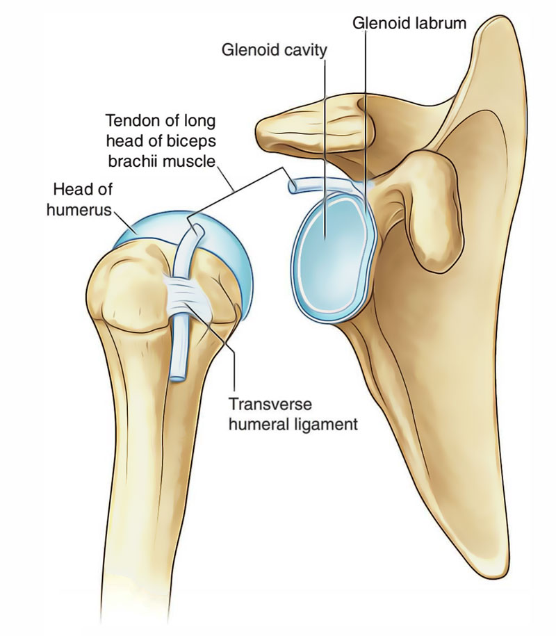
Humerus, humerus head, spatula, acetabulum, acromion, clavicle, clavivular joint, coracoid process. The shoulder joint (glenohumeral joint) is a ball and socket joint between the scapula and the humerus. Simple easy notes for quick revision for exams. You can see it enclosing the glenohumeral joint and you can see its attachment on the anatomical neck that's the shoulder joint. The next layer is made up of the ligaments of the joint capsule. Webmd's shoulder anatomy page provides an image of the parts of the shoulder and describes its function, shoulder problems, and more. The glenohumearal joint has a greater range of motion than any other joint in the body. We used a 3d volume. In human anatomy, the shoulder joint comprises the part of the body where the humerus attaches to the scapula.1 there are two kinds of cartilage in the joint. The shoulder joint is formed where the humerus (upper arm bone) fits into the scapula (shoulder blade), like a ball and socket. • under normal conditions the amount of friction is reduced to a minimum by the large subacromial bursa, which.
The human shoulder is the most mobile joint in the body. Shoulder anatomy, shoulder bone, shoulder diagram, shoulder joint bones, shoulder muscle structure, shoulder parts of the body, shoulder tendon anatomy, shoulder tendons ligaments, hand, shoulder anatomy, shoulder bone, shoulder diagram, shoulder joint bones. Just remember the articulating surfaces. The next layer is made up of the ligaments of the joint capsule.
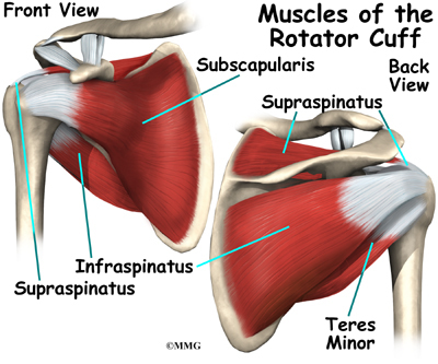
All about the shoulder muscles.
The shoulder is actually composed of four joints, namely glenohumeral joint, acromioclavicular joint, sternoclavicular joint and scapulothoracic joint. Simply put, the shoulder, or shoulder joint, is the connection of the upper arm and the thorax. Human anatomical atlas of the shoulder : Medical illustration showing deep layer of muscles, ligaments and tendos all labeled. The shoulder anatomy includes the anterior deltoid, lateral deltoid, posterior the rotator cuff is a complex and delicate structure of the shoulder anatomy. Normal anatomy, variants and checklist. Three bones come together at the shoulder joint. Describe the structure of the shoulder should begin with bone parts that include: Click now and learn everything about its anatomy and function at kenhub! All about the shoulder muscles. Axial slice of t1 weighted mri with all anatomical structures labeled. • during abduction of the shoulder joint, the supraspinatus tendon is exposed to friction against the acromion.
Joints hold the skeleton together and support movement. It is the major joint connecting the upper limb to the trunk. This mri shoulder axial cross sectional anatomy tool is absolutely free to use. The first is by joint function, also referred to as range of motion. 7 draw labelled diagram showing the relations of shoulder joint. The deepest layer of the shoulder includes the bones and the joints. You can see it enclosing the glenohumeral joint and you can see its attachment on the anatomical neck that's the shoulder joint. The small size of the glenoid fossa and the relative laxity of the joint capsule renders the joint relatively unstable and prone to subluxation and. 6 describe briefly the abduction at shoulder joint.
Just remember the articulating surfaces.
Describe the structure of the shoulder should begin with bone parts that include: Comprising of numerous ligamentous and muscular structures, the only the joint capsule attaches proximal to the glenoid fossa and attaches further distally to the anatomical neck of the humerus. Looking for quizzes, videos, articles and an. You can see it enclosing the glenohumeral joint and you can see its attachment on the anatomical neck that's the shoulder joint. Simply put, the shoulder, or shoulder joint, is the connection of the upper arm and the thorax. Learn vocabulary, terms and more with flashcards, games and other study tools. In this article, we shall look at the anatomy of the shoulder joint and its important clinical correlations. In common usage, shoulder joint mostly refers to the glenohumeral joint, the major joint of the shoulder but can also include acromioclavicular joint. In human anatomy, the shoulder joint comprises the part of the body where the humerus attaches to the scapula.1 there are two kinds of cartilage in the joint. Equally extensive are the muscles affecting the shoulder movement, including: Shoulder anatomy, shoulder bone, shoulder diagram, shoulder joint bones, shoulder muscle structure, shoulder parts of the body, shoulder tendon anatomy, shoulder tendons ligaments, hand, shoulder anatomy, shoulder bone, shoulder diagram, shoulder joint bones. This mobility provides the upper extremity with tremendous range of motion such as adduction, abduction, flexion, extension, internal rotation, external rotation, and 360° circumduction in the shoulder joint anatomy.
This diagram with labels depicts and explains the details of shoulder anatomy diagram. Equally extensive are the muscles affecting the shoulder movement, including:

Posting Komentar untuk "Shoulder Joint Anatomy Diagram"